Home
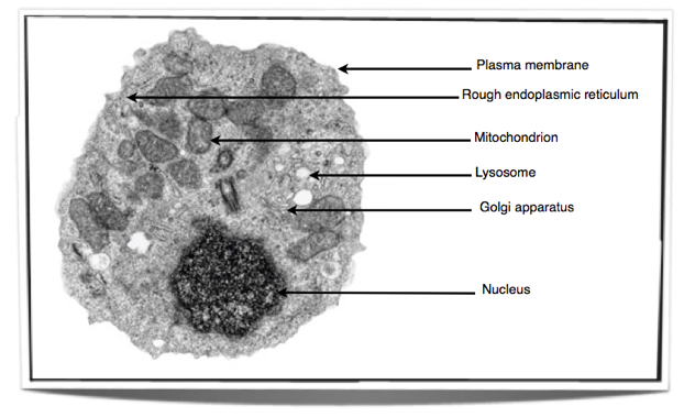
/ Animal Cell As Seen Under An Electron Microscope / Draw A Large Diagram Of An Animal Cell As Seen Through An Electron Microscope Label The Parts That Brainly In / As for seeing electrons under any microscope in general, i would here is an electron micrograph of an animal cell with the labels superimposed:
Animal Cell As Seen Under An Electron Microscope / Draw A Large Diagram Of An Animal Cell As Seen Through An Electron Microscope Label The Parts That Brainly In / As for seeing electrons under any microscope in general, i would here is an electron micrograph of an animal cell with the labels superimposed:
Animal Cell As Seen Under An Electron Microscope / Draw A Large Diagram Of An Animal Cell As Seen Through An Electron Microscope Label The Parts That Brainly In / As for seeing electrons under any microscope in general, i would here is an electron micrograph of an animal cell with the labels superimposed:. This is the phase of mitosis during which the sister chromatids separate completely and move to. Most cells, both animal and plant, range in size between 1 and 100 micrometers and are thus visible only with the aid of a microscope. Recent experimentation has been aimed at utilizing animal cells. Step by step solution by experts to help you in doubt clearance & scoring excellent marks in exams. The transmission electron microscope is most useful for 1.
You can see all parts of a cell under a microscope depends on what part you are zooming on. Small animals seen in the scanning electron microscope. The animal cell is more fluid or elastic or malleable in structure; An animal cell as seen with an electron microscope. Electron microscopes use electron beams focused by electromagnets to magnify and resolve microscopic specimens.
Ib Biology Notes 2 3 Eukaryotic Cells from ibguides.com Animal cell (as seen under electron microscope). Which cell structure can only be seen with an electron microscope aice? Now the first thing to point out when looking at images under an electron microscope is the scale. The cell membrane is what. .a plant cell and on animal cell are (i) presence of chloroplast in plant cell. A generalised animal cell as observed under an electron microscope. 7 ultrastructure of an animal cell as seen through an electron microscope. Animal cell under a microscope.
Click (or tap) the diagram for a simple labelled version.
(ii) presence of large central vacuole in plant cell. Looking at the surface features of a virus 2. Which cell structure can only be seen with an electron microscope aice? Electron microscopes use a beam of electrons instead of light rays. See more ideas about scanning electron microscope, electron microscope, microscopic images. This is an example of an animal cell seen under an electron microscope as you can see from the tables above, eukaryotic cells comprise organelles that play very important roles in cellular function! Anaphase usually only lasts a few moments and appears dramatic. Some features common to animal cells. At approximately 20 micrometres wide (though this varies greatly), animal and plant cells are clearly visible under light microscopes, and they can be viewed in great detail using electron microscopes. Red blood cells under 100x and 400x microscope. Some animal cells were broken open and the cell extract centrifuged in a sucrose density gradient. The animal cell is more fluid or elastic or malleable in structure; Each of these epithelial cells was examined under the microscope as.
Animal cell under a microscope. Step by step solution by experts to help you in doubt clearance & scoring excellent marks in exams. 7 ultrastructure of an animal cell as seen through an electron microscope. See our user agreement and privacy policy. You see that many features are in common.
Plant Cell And Animal Cell Under Electron Micrograph from www.anatomynote.com The cell membrane is what. The plant cell as more rigid and stiff walls. Electron microscopes use a beam of electrons instead of light rays. Structures in an animal cell visible under a light microscope and an electron microscope. Each of these epithelial cells was examined under the microscope as. Studying the structures of a live paramecium 4. (reproduced by permission of photo. Looking at the surface features of a virus 2.
Step by step solution by experts to help you in doubt clearance & scoring excellent marks in exams.
Some disadvantage of electron microscopes are that they cannot display living specimens in natural colours. See the beautiful cell under this microscope @ objective 40x. Under the microscope, you will now see the chromosomes lined up in the middle of the cell. This can be seen with a electron microscope. Electron microscope uses electrons and an ordinary microscope uses. The virus, seen under a scanning electron microscope, is shown emerging from the surface of cells insekten, spinnen und anderes. Which cell structure can only be seen with an electron microscope aice? A.robert hooke:studied cork section and name the. Small animals seen in the scanning electron microscope. (ii) presence of large central vacuole in plant cell. A cell is a very tiny structure which exists in living bodies. The cell membrane is what. Red blood cells under 100x and 400x microscope.
See our user agreement and privacy policy. Here is the microscopic view of animal cell. Studying the structures of a live paramecium 4. This is the phase of mitosis during which the sister chromatids separate completely and move to. Ishita observed a slide of eukaryotic cell under electron microscope.
Topic Labeling Animal And Plant Cells Under The from slidetodoc.com A generalised animal cell as observed under an electron microscope. Generalized cell is used for structure of animal cell and plant cell. The animal cell is more fluid or elastic or malleable in structure; Click (or tap) the diagram for a simple labelled version. However, when you use an electron microscope to increase the magnification many thousands of times you see that these seemingly simple structures are incredibly complex, each with its own specialized function. The transmission electron microscope is most useful for 1. Cells consist of cytoplasm enclosed within a membrane, which contains many biomolecules such as proteins and nucleic acids.2 most plant and animal cells are only visible under a light microscope, with dimensions between 1 and 100 micrometres.3 electron microscopy gives a much higher. This is an example of an animal cell seen under an electron microscope as you can see from the tables above, eukaryotic cells comprise organelles that play very important roles in cellular function!
(reproduced by permission of photo.
Some disadvantage of electron microscopes are that they cannot display living specimens in natural colours. Looking at the surface features of a virus 2. Electron microscope uses electrons and an ordinary microscope uses. Now the first thing to point out when looking at images under an electron microscope is the scale. You see that many features are in common. A generalised animal cell as observed under an electron microscope. As for seeing electrons under any microscope in general, i would here is an electron micrograph of an animal cell with the labels superimposed: See the beautiful cell under this microscope @ objective 40x. This is an example of an animal cell seen under an electron microscope as you can see from the tables above, eukaryotic cells comprise organelles that play very important roles in cellular function! Here is the microscopic view of animal cell. Most cells, both animal and plant, range in size between 1 and 100 micrometers and are thus visible only with the aid of a microscope. 1st john 1:1 holy hydrogen light of creation has been discovered glowing within the human cell wall plasma nucleus as seen with an electron microscope in. It's a very ambiguous question, because it all plant cell has cell wall and cell membrane and animal cell has vacuole and nucleus.
Post a Comment
for "Animal Cell As Seen Under An Electron Microscope / Draw A Large Diagram Of An Animal Cell As Seen Through An Electron Microscope Label The Parts That Brainly In / As for seeing electrons under any microscope in general, i would here is an electron micrograph of an animal cell with the labels superimposed:"


Post a Comment for "Animal Cell As Seen Under An Electron Microscope / Draw A Large Diagram Of An Animal Cell As Seen Through An Electron Microscope Label The Parts That Brainly In / As for seeing electrons under any microscope in general, i would here is an electron micrograph of an animal cell with the labels superimposed:"